Veterinary Radiographs (X-rays)
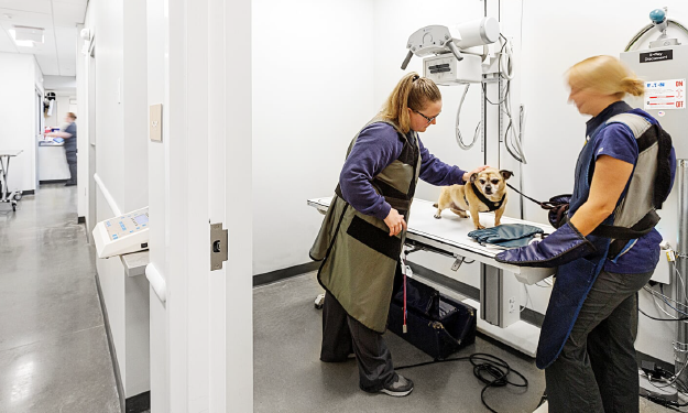
Radiographs, commonly referred to as X-rays, are a diagnostic imaging tool used to diagnose diseases and conditions in the chest, abdomen, and musculoskeletal system (bones). X-rays are the most common diagnostic tool used in veterinary medicine and are considered a safe, non-invasive tool that does not cause changes to the disease process or discomfort to the animal. Sedation or anesthesia is sometimes recommended on a case-by-case basis depending on the pet.
Radiography is employed by all the different services in veterinary medicine, from primary care veterinarians to specialty services and emergency cases. At Ethos hospitals, all x-ray images are interpreted by one of our board certified radiologists. If there is not a radiologist on-site or available, the image will be sent and interpreted by one within our network.
X-rays are used in all kinds of animals, not just dogs and cats, and are often used for rabbits, birds, and reptiles. An x-ray image can diagnose a variety of conditions in pets, including:
- Gastrointestinal obstruction (foreign bodies) & urinary obstruction
- Bone fractures and other trauma
- Respiratory distress and pneumonia
- Gastric Dilatation Volvulus (GDV) and bloat
- Pregnancy
- Orthopedic conditions (hip dysplasia, arthritis, & more)
- Dental disease
- Cancer
- Many, many other conditions
Click any of the images below to enlarge them
-
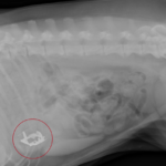
- X-ray of a dog that ingested a toy car.
-
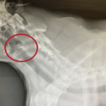
- X-ray of a dog that swallowed a fish hook.
-
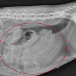
- X-ray of a cat pregnant with two kittens.
-
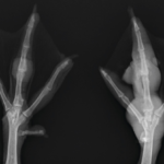
- X-ray of a duck with bumble foot.
-
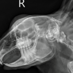
- X-ray of a rabbit in need of a tooth trim.
-
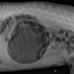
- X-ray of a dog with bloat.
-
- X-ray of a dog with healed acetabular fractures (broken hip socket – left side) sustained after being hit by a car.
-
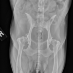
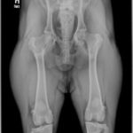
- X-ray of a dog with severe hip dysplasia.