Tetatnus in Dogs: A Case Study
Within a 24 hour period in April 2021, two dogs presented to Capital District Veterinary Referral Hospital showing signs of tetanus. Being such a rare condition, these cases are a good review of the disease and treatment.
Presentation:
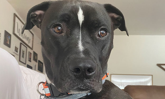
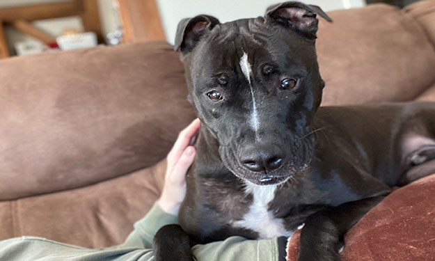
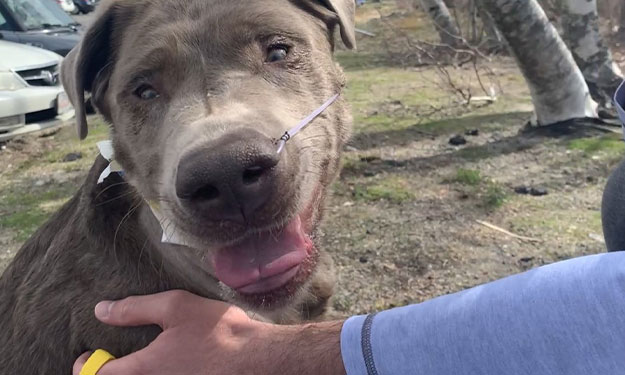
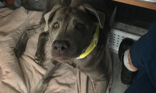
Floki, a 2 year old CM Pitbull, presented to our emergency room as a referral from a local veterinarian for presumed tetanus. Not hours prior, we had sent another dog, Bear, a 2 year old CM Labrador Retriever, to Massachusetts Veterinary Referral Hospital for continued care including tetanus antitoxin and possible ventilation.
History and Physical Examination:
Interestingly, both dogs had a nail bed infection of the 4th digit of the left forelimb. Floki’s nail was cut too short by the owner 6 days prior and Bear had broken his toenail on a walk in the woods 10 days before. Both nail beds had sealed over and there was no inflammation, swelling, or discharge visible on initial examination. The only way we knew of the injuries was from the owner’s history.
Both dogs exhibited ocular and facial abnormalities as their first clinical signs. Then over 24-48 hours, signs progressed rapidly to rigidity of localized muscles to generalized spastic paralysis including contracted facial musculature, trismus, retracted lips, wrinkled forehead, rictus grin, miosis, enophthalmos, prolapse of the third eyelid, erect ears, and hyperextension of the limbs and stiff gait. Both dogs could still walk and eat, thankfully.
Differential diagnoses:
The physical examination changes are so unique, it is hard to mistake tetanus for another disease process, however, if the dogs are showing only mild signs, the differential diagnosis include polymyositis, strychnine intoxication, spinal trauma, hypocalcemia, and meningoencephalitis.
Diagnosis:
Based on the physical exams of both dogs and their recent wounds, they were diagnosed with tetanus. Tetanus is caused by the neurotoxin, tetanospasmin (TeNT), produced by Clostridium tetani. This anaerobic bacteria is found in the soil all over the world, however, germination only occurs with temperatures >20°C (68°F) and humidity reaching at least 15%.
If the wound is deep, necrotic, and not open to the air (anaerobic), then the bacterium can sporulate and release the toxin. Tetanus toxin diffuses locally and interacts with nerve endings. The toxin undergoes retrograde transport to the neuronal cell bodies in the central nervous system where it blocks the release of glycine and GABA. This inhibits the inhibitory effects on the motor neuron thereby increasing firing of the motor neurons and causing muscle spasms.
Clinical signs:
The characteristic clinical signs are caused by this muscle contracture and spasming. The most common are marked muscle rigidity and altered facial expression (signs listed above). Other signs may include dysphagia and ptyalism, hyperesthesia and ataxia. Death can occur from respiratory arrest secondary to either rigidity of external respiratory musculature, laryngospasm, increased airway secretions or even central respiratory arrest. Markedly affected patients who cannot walk and are down may develop respiratory or urinary infections and even decubitus ulcers if not cared for properly.
Treatment:
There is no specific treatment or reversal/antidote for tetanus. Standard care includes injection of a tetanus antitoxin (TAT) which is an equine serum taken from horses inoculated with C. tetani. It is only effective against the circulating/unbound toxin, so it can help dogs from getting worse but cannot reverse toxin already bound to CNS. It also cannot cross the blood brain barrier to remove unbound toxin in the CNS. Interestingly, accordingly to veterinary studies on tetanus, which are limited, there is not any difference in survival between patents who received TAT and those who did not. The recommended dose is 100-10,000 units/kg subcutaneously. Side effects include anaphylaxis and serum sickness. We did not have tetanus antitoxin at CDVRH and therefore, Bear was referred to Massachusetts Veterinary Referral Hospital for continued care.
Goals of treatment are based on severity of signs; some dogs need sedation and muscle relaxation and some require a mechanical ventilator. All animals are treated with an antibiotic to kill off Clostridium such as a penicillin or metronidazole but this does nothing for the disease itself since the released toxin has bound to the neurons, however, it prevents more toxin from being produced.
The most important part of this treatment is evaluating the wound. As a veterinary professional reading this, you would think, ‘of course’, but I want this article to emphasize this point. I evaluated the nail bed of Bear and deemed it healed. There was no swelling, pain, or discharge. It was not even red. Then, after I received advice from a neurologist, I re-evaluated it under heavy sedation and opened up the healed nail bed. It felt weird cutting into healthy tissue, but under it, I found purulent material at the nail bed and actually traveling down the nail itself. I flushed out the purulent exudate, cut the nail, and soaked the foot in hydrogen peroxide to give this anaerobic bacterium more oxygen.
It is important to remember that these dogs should be kept in a quiet, dark room. We placed cotton balls in the dog’s ears, covered their cages with blankets, and placed them in a quiet area. Some dogs need intravenous fluids to maintain hydration and feeding tubes for enteral nutrition if unable to eat. Muscle relaxants and sedation, such as methocarbamol 30-44mg/kg IV or PO q 8 or midazolam/diazepam 0.2 – 0.5mg/kg IV PRN, Acepromazine 0.01-0.05mg/kg IV PRN, and/or Trazodone 5-7mg/kg PO q 8, are very beneficial. Magnesium sulfate can be used as muscle relaxation as well since it can block calcium’s entry to the neuromuscular junction. For our patients, we mainly used methocarbamol and acepromazine.

Outcomes:
I received updates (and photos) from the owners of both dogs and thankfully, both, are doing great. It took Floki 7 weeks to return to normal but was able to do this at home under care from the owners. Bear stayed at Massachusetts Veterinary Referral Hospital for 2 weeks and required amputation of the infected digit and a nasogastric feeding tube due to progression of dysphagia but did not require the mechanical ventilator.
References
- Tetanus in animals. J Vet Diagn Invest. March 2020;32(2):184-191. Michel R Popoff
- Lecture: Tetanus in Dogs and Cats from Veterinary Emergency and Critical Care 2019 Spring Symposium by Adesola Odunayo, DVM, MS, DACVECC