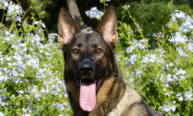
Case Study: Canine Foreign Body

Written by Joshua Jackson, DVM, Diplomate ACVS
History:
Jake, a 2.5 year old German Shepherd, was presented for evaluation of vomiting, anorexia and lethargy. Prior to referral, Jake had a 5 month history of vomiting, diarrhea, weight loss and inappetence. He was treated supportively with a bland diet, maropitant and omeprazole. Although his vomiting ceased and he began eating, Jake remained persistently lethargic. He was evaluated five days after initiation of supportive care. A leukocytosis (17.4K) characterized by a monocytosis and thrombocytopenia (98K) were revealed. A spec PLI was normal. Supportive care was continued and Jake apparently improved over the next few days. His signs did not completely resolve, thus he was presented for further evaluation.
Physical Examination Findings:
Bright, Alert and Responsive
Body condition score 3/9
Very tense on abdominal palpation, but all other parameters and vitals normal.
Differential Diagnoses:
Primary gastrointestinal disease including gastroenteritis vs. partial obstruction (foreign body) vs. intussusception vs. mesenteric or splenic torsion vs. less likely parasitism vs. secondary to endocrine disease such as hypoadrenocorticism. Renal, hepatic and pancreatic disease are less given results of biochemical analysis and cPLI.
Diagnostics:
Abdominal ultrasound: A linear foreign body within the peritoneal cavity with a diffuse peritonitis was evident. Regional intestinal mural alterations, likely secondary to inflammation or adhesion formation, were noted, as well as periaortic lymphomegaly and hypoechoic hepatomegaly.
Plan:
Exploratory celiotomy and foreign body removal.
Surgery Report:
A standard ventral midline incision was made from xiphoid to pubis. 300mls of purulent ascites was encountered upon entry into the abdomen. Diffuse omental and intestinal adhesions were present. A large toothpick foreign body was discovered floating free in the ascites. Adhesions were removed and copious saline lavage performed. A closed suction drain was placed. Closure was routine.
Discussion:
Chronic peritonitis, especially septic peritonitis is not common. Animals are able to tolerate severe abdominal inflammation and hide their clinical signs much more than people. Historical veterinary literature suggests a mortality rate of septic peritonitis from 25-75%. It is our experience that with aggressive medical and surgical management survival rates are much better than what has been reported historically.
Open peritoneal drainage is almost never performed and almost all patients are managed with closed suction drainage. One of the most important components of successful postoperative management of the peritonitis patient is addressing the often severe hypoproteinemia. Early post operative enteral feeding is important. The use of nasogastric, esophagostomy, gastrostomy or jejunostomy tubes should be considered standard of care with septic peritonitis. Colloid support in addition (hetastarch, plasma, albumin) is often required. Patients often lose tremendous volumes of fluid from the abdomen. A close watch of blood pressure, body weight and hydration status is important and volume administration adjusted accordingly. The use of paired blood and abdominal fluid glucose levels has not been validated in the post operative period to determine if sepsis is present, but it is our experience that a lower abdominal glucose than plasma in the post operative period generally suggests recurrent leakage. A closed suction drain enables easy access for cytology to evaluate WBCs morphology and presence of bacteria. Broad spectrum antibiotic therapy is generally utilized. We often utilize enrofloxacin combined with ampicillin or ticarcillin.
In this case, the patient was discharged 3 days after surgery and has done well in the postoperative period.