Early Orthodontic Intervention in Veterinary Dentistry
February 11, 2026
National Pet Dental Health Month provides an opportunity to reflect on how advances in veterinary dentistry are shaping patient care, particularly when conditions are identified and addressed early. Across the Ethos Veterinary Health network, specialty dental teams apply evidence-based approaches that prioritize comfort, function, and long-term oral health.
One such team is based at Animal Dental Center (ADC), an Ethos network hospital known for its work in complex dental and maxillofacial cases, including trauma and oral oncology. Led by Mary Krakowski Volker, DVM, DAVDC, ADC functions as a referral center for advanced dental care.
The following case demonstrates how early orthodontic intervention is applied in clinical practice.
A comfortable, functional occlusion allows dogs and cats to open and close their mouths and chew without pain or trauma from opposing dentition. In veterinary dentistry, orthodontic intervention is used to treat traumatic malocclusions and achieve a pain-free, functional, and stable occlusion. Treating permanent teeth during active eruption allows for faster and more predictable results. Occlusion should be monitored during juvenile examinations, and when a malocclusion is identified, timely intervention is recommended. Maxillary and mandibular canine teeth are most commonly treated due to their size and potential to cause occlusal trauma when malpositioned.
The case presented here utilizes multiple orthodontic techniques, including one active force appliance and two passive force appliances, demonstrating prompt intervention during eruption of permanent canine teeth. Orthodontic treatment typically involves multiple anesthetic events (2 to occasionally 4) over a four- to eight-week period, with outcomes dependent on staged intervention and post-procedural management.
Case Presentation
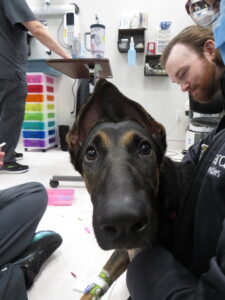
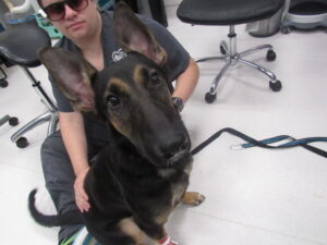
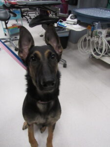
“Mavis”
Five-month-old, female, GSD
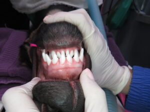
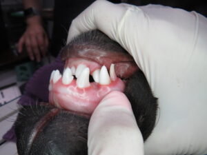
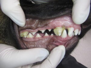
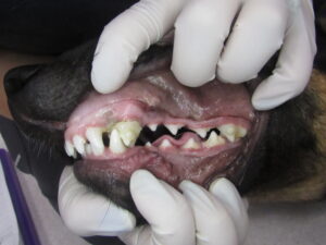
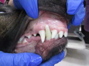
Mavis presented with persistent deciduous maxillary canine teeth (504, 604) and a mild Class II malocclusion characterized by short mandibles, linguoverted (base-narrow) mandibular canines (304, 404), and mesioverted (lance) maxillary canines (104, 204).
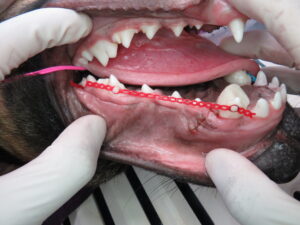
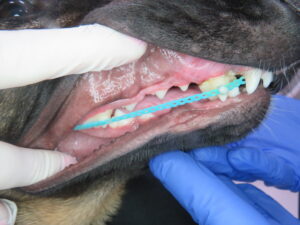
Treatment Day 1
 Persistent deciduous maxillary canines (504, 604) were extracted. Masel chains were placed on the maxillary canine teeth (104, 204) and anchored to the maxillary fourth premolar (108, 208) and first molar (109, 209). These chains encouraged distal tipping of the maxillary canine teeth to widen the diastema and create space for the crowns of the mandibular canine teeth (304, 404).
Concurrently, incline planes connected to only the maxillary canine teeth (104, 204) were placed to tip the mandibular canine teeth (304, 404) buccally. The combined pressure of the 304, 404 on the incline planes attached to the 104, 204, may also create enough strategic pressure to provide bodily movement of the 104, 204 distally.
Treatment Day 7
Mavis had a conscious (awake) recheck examination 1 week after placement of her orthodontic appliances. The appliances were creating appropriate tipping movement of the 104, 204, 304, 404.
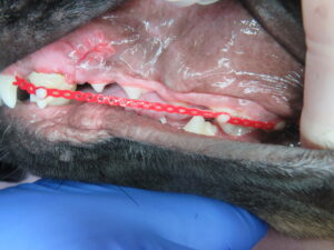
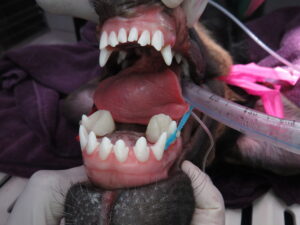
Treatment Day 17
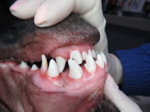
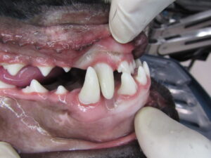
Approximately two and a half weeks following appliance placement, the mandibular canines have tipped appropriately buccally and the maxillary canine teeth tipped distally – there is now a diastema present for the cusps of the mandibular canine teeth. The canine teeth are ~50% erupted.
The appliances (orthodontic buttons, masel chains, and incline planes) are removed. The canine teeth are not erupted yet to achieve retention (i.e. the 304, 404 cusps are not yet erupted enough to sit appropriately buccally in the maxillary diastema bilaterally), thus acrylic crown extensions are placed on the 304, 404 crowns to lengthen their cusps and retain their tipped position.
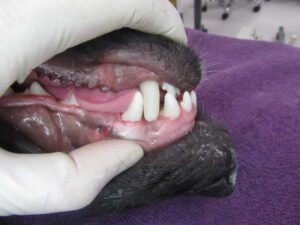
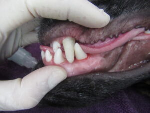
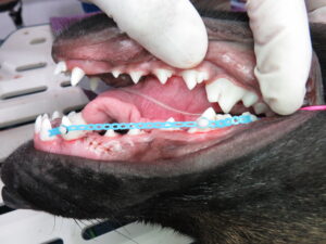
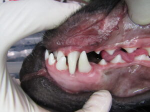
Treatment Day 31
Four weeks post orthodontic treatment, the canine teeth have now almost completely erupted. The crown extensions are reduced to a small (~2-3 mm) amount of extension left to ensure retention as the canine teeth finish erupting. This small amount of extension can be removed at Mavis’ first COHAT around 12-16 months of age. Mavis now has a comfortable, functional occlusion, that will remain stable for her lifetime!

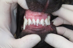
A post orthodontic treatment recheck was performed using a conscious oral examination. Tooth positioning and occlusion remained appropriate, confirming long-term treatment success.


Clinical Takeaway
Early orthodontic intervention during active tooth eruption allows for predictable tooth movement and functional occlusal outcomes. This case highlights the role of early assessment, appropriate case selection, and staged treatment planning in veterinary orthodontics.
We’re proud of the ADC team and their work, which reflects our shared commitment to advancing specialty medicine through thoughtful clinical judgment, collaboration, and education.