What Actually Happens During Cataract Surgery?
February 20, 2016Welcome to the second article in a series about the management of diabetic cataracts in the dog. This installment deals with the cataract surgery itself. The surgical description applies to cataract surgery for any patient, diabetic or not.
For most patients, cataract surgery can be an outpatient procedure for which they are dropped off in the morning and go home in the evening. My observation is that diabetic dogs have a better chance of eating a normal meal in the evening if they are at home, which is an additional benefit to outpatient surgery.
The typical day of a cataract patient begins after drop-off with several rounds of eye drops, placement of an IV catheter, and a first blood glucose. This is to determine glucose levels as families are instructed to withhold morning food and give ½ the usual dose of insulin. We continue careful monitoring of blood glucose throughout the day with more than one measurement while under anesthesia and during recovery (which takes place in our Intensive Care Unit for all cataract surgery patients), and care is taken to keep blood glucose in a safe range throughout the day with treatments given on an as-needed basis.
The surgery is performed with the patient in dorsal recumbence, head ventroflexed, and positioned so that the eyes are level. We use special endotracheal tubes to ensure the airway is protected with the neck in ventroflexion. The animal is under general anesthesia and on a ventilator so that atracurium (a neuromuscular inhibitor) can be administered to keep the eyes central rather than rotated downward. For maximum safety, in addition to using a ventilator, the effect of atracurium is carefully monitored using a peripheral nerve stimulator.

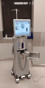
Phacoemulsification Machine
The surgery itself is performed under an operating microscope. First, an incision is made in the cornea, and the anterior chamber of the eye is entered. A substance called viscoelastic is injected into the anterior chamber to help it keep its shape during the procedure. A stab incision is made in the anterior lens capsule, and from it, a circular hole is created, exposing the lens fibers that are to be removed by phacoemulsification. The needle of the phacoemulsification unit is inserted into the lens fibers, and they are simultaneously broken up by ultrasound and vacuumed from the eye. During the process, grooves are made in the lens nucleus, and it is eventually broken into pieces which are pulverized and removed. The machine uses advanced fluidic technology of balanced salt solution flowing into and out of the eye to minimize stress on the eye during the process. All lens fibers from both the lens nucleus and cortex are removed, and the empty lens capsule is then “polished” to make it ready to accept the replacement lens. The replacement lenses are made especially for dogs with the appropriate diopter strength to restore normal vision. The viscoelastic is removed from the eye at the end of the procedure, and the corneal incision is closed with 9-0 vicryl.
Anesthetic recovery and post-operative care take place in our ICU. Short-term adjustments in diabetic control are made if needed, and there are hourly eye drops given during the afternoon. The dogs are visual at the point they wake up from anesthesia.