CT Scan for Pets: What You Need to Know
October 1, 2016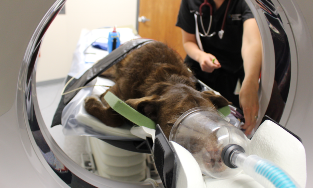
Computed Tomography (CT), commonly referred to as a Cat Scan or CT, uses x-rays and a computer to generate cross-sectional images of a region of interest. In veterinary medicine, a CT scan is often used for imaging of the chest, abdomen, nose, bones and joints.
How does a CT scan work?
Computed Tomography is commonly referred to as a CT scan. This technology is very useful when looking at parts of the body such as the lungs and thorax, nasal passage and sinuses, ears, abdomen and some orthopedic structures. CT scans are non-invasive and are not painful.
The table on which the animal is laying is slowly advanced into the part of the machine that performs the scan (called the gantry). An x-ray tube rotates 360° around the patient to record the x-rays from many angles, creating slices. The number of images taken depends on the area and size of the suspected problem. When the computer finishes processing the information, the slices are stacked together to create a three dimensional image of your pet without superimposition of organs or other tissues.
Why has a CT scan been recommended for my pet?
Animals who are candidates for a CT scan include those with:
- Lung disease
- Metastatic cancer prior to surgery
- Pulmonary fibrosis
- Masses or tumors within the chest cavity
- Nasal cavity disease (tumors vs. inflammation/rhinitis)
- Development orthopedic disease (elbow dysplasia, osteochondritis dessicans)
- Spinal or pelvic trauma (i.e. following hit-by-car accidents)
- Vascular Anomalies – (i.e. portosystemic shunts)
CT scans provide better differentiation of bones and soft tissues than conventional radiographs (x-rays) because this slice-based x-ray technology avoids the superimposition of structures that occurs with radiology, and because CT can differentiate between fluid and soft tissue. An iodine-based contrast media is usually injected intravenously to further increase the differentiation of tissue. The results of the CT scan will help your veterinarian make a definitive diagnosis and offer you the best options for treating your pet.
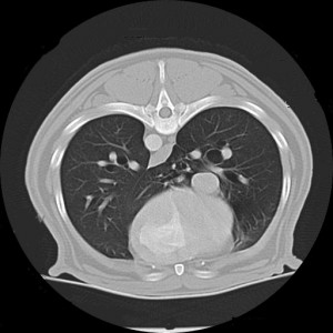
Normal examination of the lungs with thoracic CT.
Are there any known complications from a CT scan?
CT scans are considered safe, but they do utilize ionizing radiation (like an x-ray). There can be some side effects associated with the iodine-containing contrast which may be injected during the procedure. If your pet has seizures, kidney disease, or cardiac disease, we may elect to use a different type of contrast or not give contrast at all. If CT is used to guide tissue sampling, side effects can include bleeding or a leak of the lung which leads to air around the lung.
How should I prepare my pet for the CT scan?
Pets having a CT scan must be anesthetized so that they remain still for the procedure, which lasts about 30-60 minutes. In preparation for general anesthesia, please follow the instructions provided by your veterinarian for fasting. Ask your veterinarian for instructions if your pet is on any medications.
What should I expect during my pet’s CT scan?
Your pet will be anesthetized approximately 30-60 minutes for the CT scan. Before any anesthesia is given, we will make sure that your pet is healthy enough to undergo anesthesia. We will place an intravenous catheter for fluid administration throughout the procedure. Your pet’s vital signs will be carefully monitored during and after anesthesia.
What happens to my pet after the CT scan?
After the CT scan, we will watch your pet closely until he/she has recovered. Once your pet is standing and able to move around safely, he/she will be able to be discharged to you. You will be notified as to when you may pick up your pet.
Your pet may urinate a large amount after they get home because of the fluids given during the anesthesia. Once home, it will be important to keep your pet away from stairs and obstacles and feed only a small meal. The effects of general anesthesia should be gone within about 24 hours. If you feel your pet has not fully recovered by that time, please call us or your regular veterinarian.
How will I learn the results of the CT scan?
A board certified veterinary radiologist will review and create a report based on your pet’s CT images. Then, the specialist who recommended and performed your pet’s procedure will contact you and your veterinarian.
What will my pet experience during a CT scan?
We made a video giving an inside look at a veterinary CT machine being used on a dog.