Lateral Suture Repair in Canine Cruciate Ligament Surgery
March 31, 2018
Canine Cruciate Ligament Injury (CCLI) is the leading cause of pelvic limb lameness in dogs. Although tearing of this ligament can occur as a result of an athletic injury, a significant number of patients suffer from degeneration of the ligament that leads to failure with minimal trauma. The primary cause of this degeneration is unknown but likely due to several factors, including genetics and conformation. Veterinary surgeons and researchers constantly study potential contributing factors to CCLI and explore surgical techniques that may improve long-term function.
Diagnosis
The diagnosis of CCLI can be challenging for general practice veterinarians and veterinary surgeons alike. The combination of training, experience, and collaborative approach of the surgical specialists at Veterinary Specialty Hospital ensures an accurate diagnosis of CCLI despite the various clinical presentations. For complex cases, advanced diagnostic imaging techniques, including magnetic resonance imaging (MRI), computed tomography (CT), or diagnostic arthroscopy, are available in-house to help differentiate CCLI from other injuries of the stifle joint (knee). In the vast majority of clinical cases, the orthopedic exam and radiographic findings are sufficient to make a diagnosis of cruciate injury.
Treatment
The board certified surgeons at the Veterinary Specialty Hospital are trained in several surgical techniques for the repair of CCLI in dogs including
- Lateral Suture
- Tibial Plateau Leveling Osteotomy (TPLO)
- Tibial Tuberosity Advancement (TTA)
In general, surgical repair either involves the implantation of a prosthesis to mimic the function of the cruciate ligament or alteration to the stifle joint geometry to eliminate the need for the cruciate ligament. Your surgeon will consider several factors when making recommendations for surgical intervention for your pet. These include age, weight, activity, body confirmation, limb confirmation, and concurrent illness or disease. While all of the surgical techniques are likely to restore pain-free limb function, each procedure may offer specific advantages for your dog.

Lateral Suture
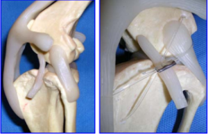
The Lateral Suture technique (lateral fabellotibial prosthesis) was developed in the early 1960s and was once considered the “gold standard” for cruciate repair in dogs. The procedure involves the placement of a prosthesis (generally a synthetic, heavy gauge suture material) in a plane parallel to the cruciate ligament. When placed correctly, this suture helps restore stability to the stifle joint (knee) by preventing the tibia from sliding forward with respect to the femur. The technique also prevents excessive internal rotation of the tibia. The scar tissue that forms around the implant further provides joint stability.

Tibial Plateau Leveling Osteotomy (TPLO)
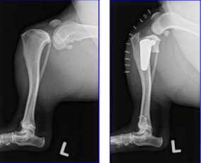
The Tibial Plateau Leveling Osteotomy (TPLO) is a novel technique that changes the weight-bearing surface of the tibia (the tibial plateau). The weight-bearing surface is altered by creating a controlled fracture (osteotomy) and rotating the tibial plateau to a more neutral or level position. A bone plate and screws stabilize the osteotomy until healing is complete. Altering the weight-bearing surface of this joint eliminates the need for the cruciate ligament. Research has shown this surgical technique to slow and potentially halt the progression of arthritis.

Tibial Tuberosity Advancement (TTA)
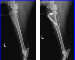
The Tibial Tuberosity Advancement (TTA) is a technique developed in Switzerland between 2002-2004. Like the TPLO, this technique involves the creation of a controlled fracture (osteotomy) to alter the geometry of the stifle joint to restore stability. This procedure also utilizes surgical implants to stabilize the osteotomy until bone healing is complete. This technique has the potential advantage of avoiding the bone’s weight-bearing surface, which may result in a more rapid return to normal function.
Prognosis
In general, the prognosis for the return to pain-free function following surgery is very high. Following an initial 2-3 week period of extreme exercise restriction, a gradual increase in controlled activity is permitted. Your surgeon will review specific recommendations regarding postoperative rehabilitation during the initial consultation and follow-up visits. Postoperative physical rehabilitation, in some cases, can maximize the positive outcome and shorten the rehabilitation period. Fortunately, the majority of dogs do not require additional physical therapy beyond leash-controlled activity at home. Potential complications exist for any orthopedic surgery and include infection, implant failure and incision complications. An experienced surgeon and appropriate postoperative care significantly decrease the risk and negative effects of these complications.